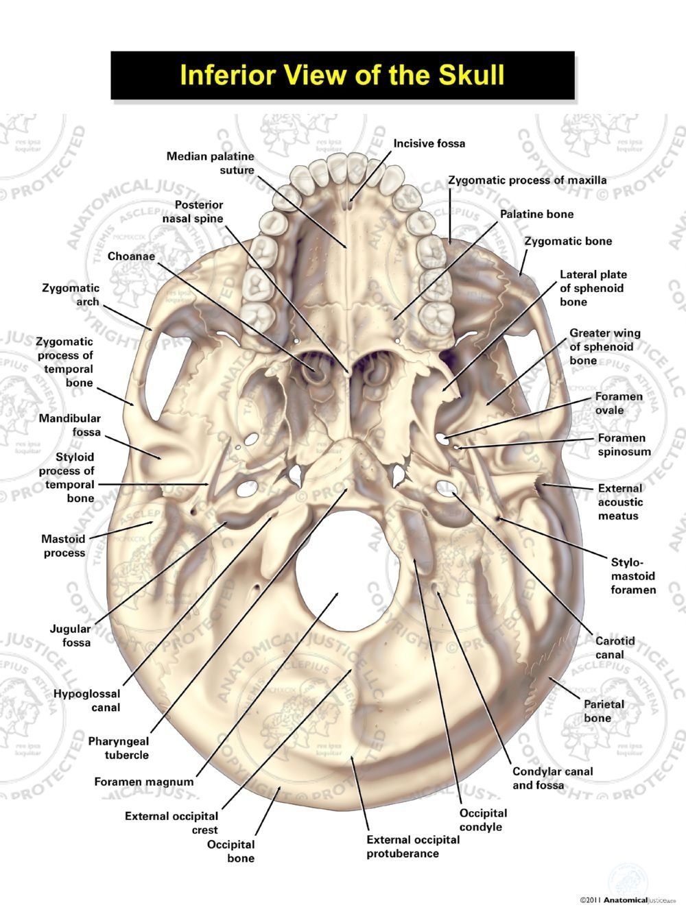
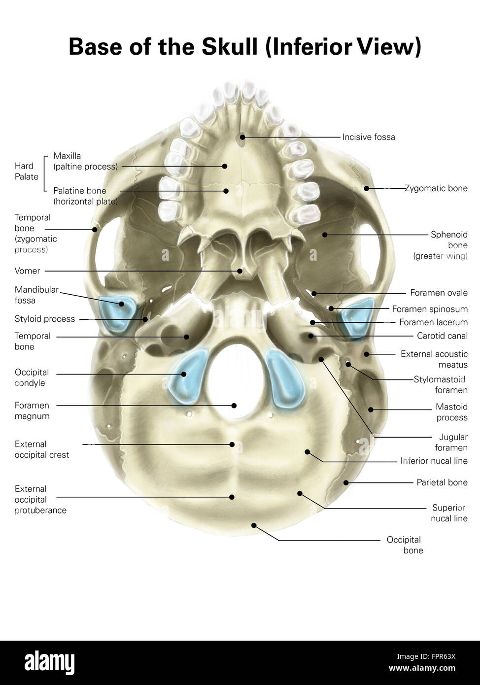
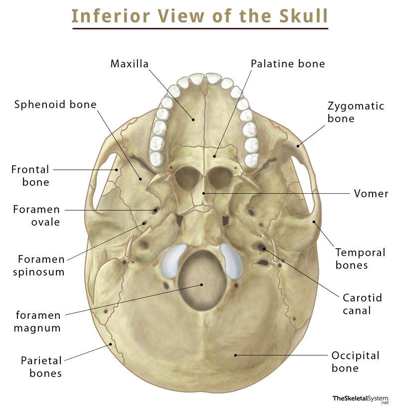
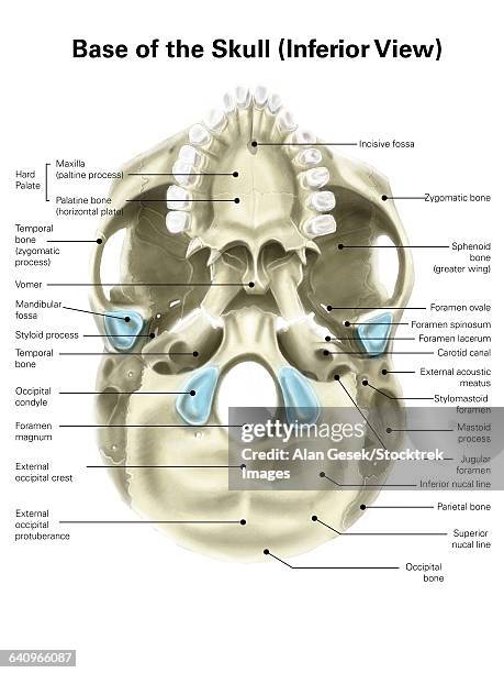
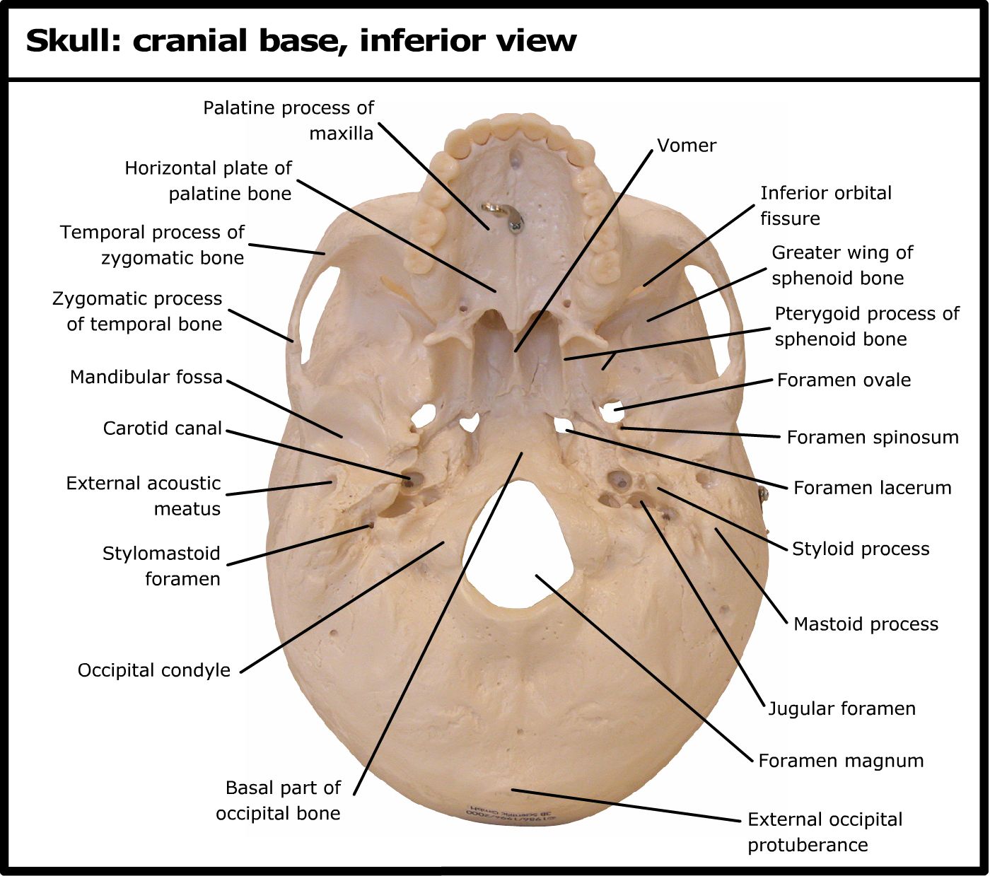
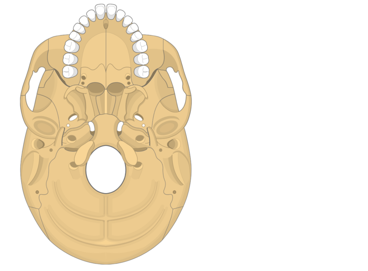
38 inferior skull anatomy labeled
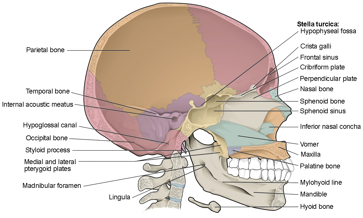
The Skull | Anatomy and Physiology I - Lumen Learning On the inferior aspect of the skull, each half of the sphenoid bone forms two thin, vertically oriented bony plates. These are the medial pterygoid plate and lateral pterygoid plate (pterygoid = "wing-shaped"). The right and left medial pterygoid plates form the posterior, lateral walls of the nasal cavity. Skull: Anatomy, structure, bones, quizzes | Kenhub The skull base is the inferior portion of the neurocranium. Looking at it from the inside it can be subdivided into the anterior, middle and posterior cranial fossae. The skull base comprises parts of the frontal, ethmoid, sphenoid, occipital and temporal bones.
1.2: Anatomical Position and Planes - Biology LibreTexts Anatomical position for a human is when the human stands up, faces forward, has arms extended, and has palms facing out. Figure 1.2. 1 : These two people are both in anatomical position. (CC-BY, Open Stax ) When referencing a structure that is on one side of the body or the other, we use the terms "anatomical right" and "anatomical left.".
Inferior skull anatomy labeled
Skull | Definition, Anatomy, & Function | Britannica The facial area includes the zygomatic, or malar, bones (cheekbones), which join with the temporal and maxillary bones to form the zygomatic arch below the eye socket; the palatine bone; and the maxillary, or upper jaw, bones. The nasal cavity is formed by the vomer and the nasal, lachrymal, and turbinate bones. Skull: cranial base, inferior view - University of Colorado Boulder Back to Model Index Page Temporal bone: anatomy and labeled diagram | GetBodySmart Carotid canal (canalis caroticus tem-poralis) is a prominent hole on the inferior surface of petrous part of the temporal bone, just anterior to jugular foramen. It gives passage for the internal carotid artery to enter the base of the skull. The internal carotid artery then curves anteromedially and enters the cranium through the foramen lacerum.
Inferior skull anatomy labeled. 8.2.3: Markings of the Cranium - Biology LibreTexts Markings of the Cranium Attributions (All Skull Sections) Markings of the Cranium Recall from Chapter 7: Introduction to the Skeletal System, that bones have markings including holes, passageways, basins, and projections. Inferior view of the base of the skull: Anatomy | Kenhub It is an unpaired bone that forms the posterior inferior part of the bony nasal septum. The sphenoid bone sits within the centre of the skull base like a wedge. This bone articulates with the vomer inferiorly, and the greater wings extend laterally to form part of the anterior pterion joint. The Skull Bones Anatomy - Inferior View | GetBodySmart Let's start with taking a look at the cranial and facial bones from an anterior view before we dive into their markings from an inferior perspective. Facial Bones: Zygomatic bone ( os zygomaticum ). Maxilla bone ( os maxilla ). Palatine bone ( os palatinum ). Learn skull anatomy faster with these interactive skull bones quizzes and worksheets. Inferior Skull Labeling Quiz - PurposeGames.com February 22, 2022 - 12:00 am There is a printable worksheet available for download here so you can take the quiz with pen and paper. From the quiz author Label the superficial markings and bones of the inferior skull. Remaining 0 Correct 0 Wrong 0 Press play! 0% 0:00.0 Show More Other Games of Interest Nail Structure Science English Creator
7.3 The Skull - Anatomy & Physiology On the inferior aspect of the skull, each half of the sphenoid bone forms two thin, vertically oriented bony plates. These are the medial pterygoid plate and lateral pterygoid plate (pterygoid = "wing-shaped"). The right and left medial pterygoid plates form the posterior, lateral walls of the nasal cavity. The Skull: Names of Bones in the Head, with Anatomy, & Labeled Diagram Inferior View of the Skull Blood Supply The skull primarily gets its blood supply from the common carotid artery, while the vertebral artery also contributes. Muscles The scalp and face muscles are innervated mainly by the facial, oculomotor, or trigeminal nerves. The hypoglossal nerve innervates the tongue. Skull Labeling - Inferior view Flashcards | Quizlet Skull Labeling - Inferior view 5.0 (1 review) Term 1 / 15 zygomatic bone Click the card to flip 👆 Definition 1 / 15 Click the card to flip 👆 Flashcards Learn Test Match Created by ryanjmartin98 Terms in this set (15) zygomatic bone sphenoid bone vomer zygomatic process of temporal bone styloid process mastoid process occipital condyle temporal bone 7.2 The Skull - Anatomy and Physiology 2e | OpenStax On the inferior aspect of the skull, each half of the sphenoid bone forms two thin, vertically oriented bony plates. These are the medial pterygoid plate and lateral pterygoid plate (pterygoid = "wing-shaped"). The right and left medial pterygoid plates form the posterior, lateral walls of the nasal cavity.
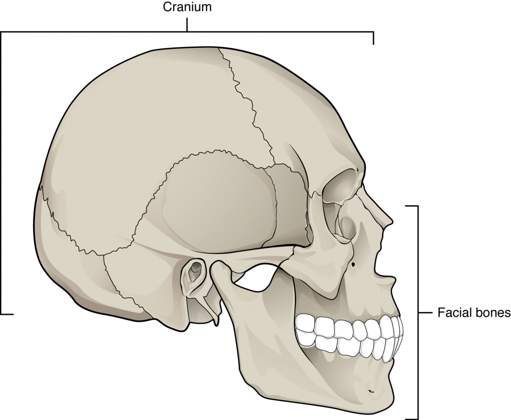
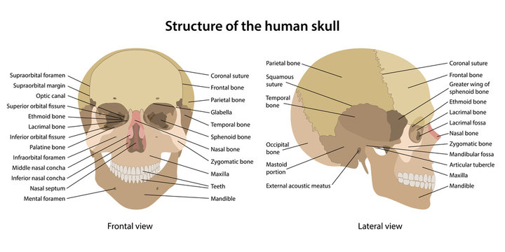
Skeletal System - Labeled Diagrams of the Human Skeleton - Innerbody The bones of the superior portion of the skull are known as the cranium and protect the brain from damage. The bones of the inferior and anterior portion of the skull are known as facial bones and support the eyes, nose, and mouth. Hyoid and Auditory Ossicles. The hyoid is a small, U-shaped bone found just inferior to the mandible. The hyoid is ... A 3D stereotactic atlas of the adult human skull base The skull base region is anatomically complex and poses surgical challenges. Although many textbooks describe this region illustrated well with drawings, scans and photographs, a complete, 3D, electronic, interactive, realistic, fully segmented and labeled, and stereotactic atlas of the skull base has not yet been built. Our goal is to create a 3D electronic atlas of the adult human skull base ... Skull - Knowledge @ AMBOSS Adult skull. The calvaria comprises the superior portions of the frontal bone, the occipital bone, and the parietal bones.. Infant skull (fontanelles). An infant's neurocranium consists of five separate bones (two frontal bones, two parietal bones, and one occipital bone) held together by connective tissue sutures. This allows for stretching and deformation of the skull to facilitate birth and ... Bones of the Skull - Structure - Fractures - TeachMeAnatomy It is comprised of many bones, which are formed by intramembranous ossification, and joined by sutures (fibrous joints). The bones of the skull can be considered as two groups: those of the cranium (which consist of the cranial roof and cranial base) and those of the face.
Temporal bone: anatomy and labeled diagram | GetBodySmart Carotid canal (canalis caroticus tem-poralis) is a prominent hole on the inferior surface of petrous part of the temporal bone, just anterior to jugular foramen. It gives passage for the internal carotid artery to enter the base of the skull. The internal carotid artery then curves anteromedially and enters the cranium through the foramen lacerum.
Skull: cranial base, inferior view - University of Colorado Boulder Back to Model Index Page
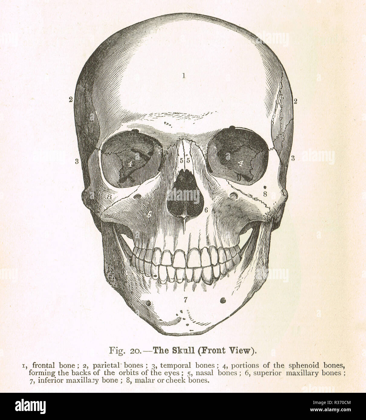
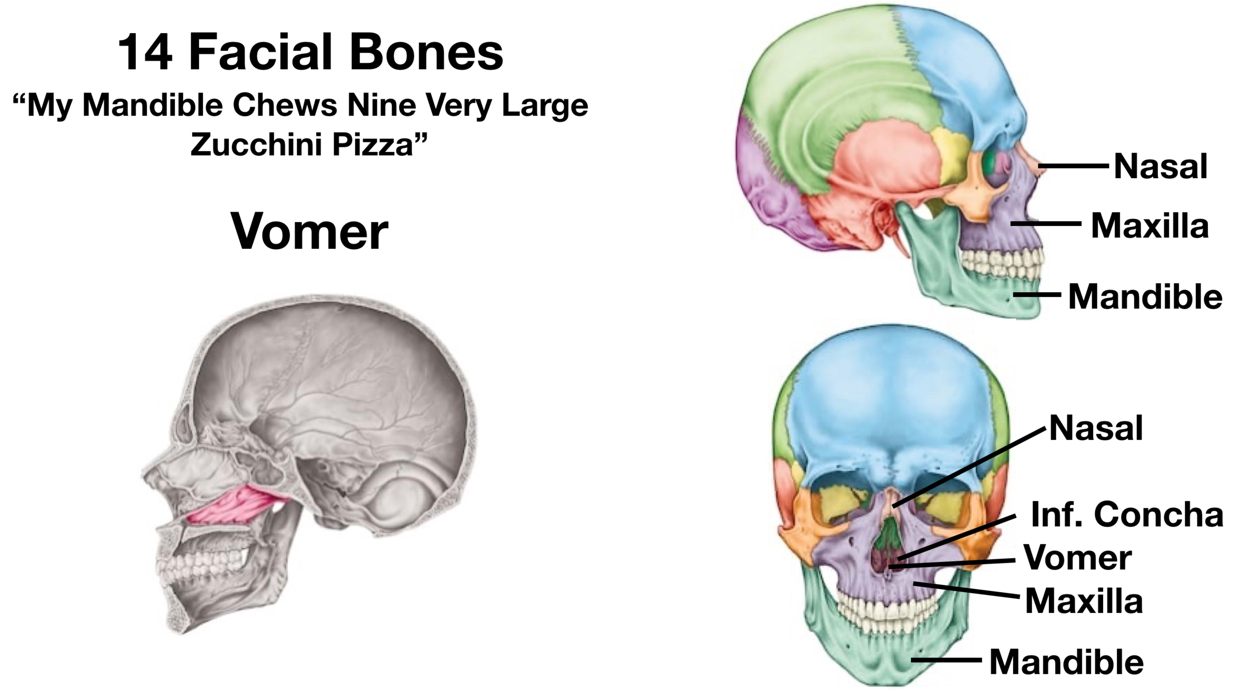
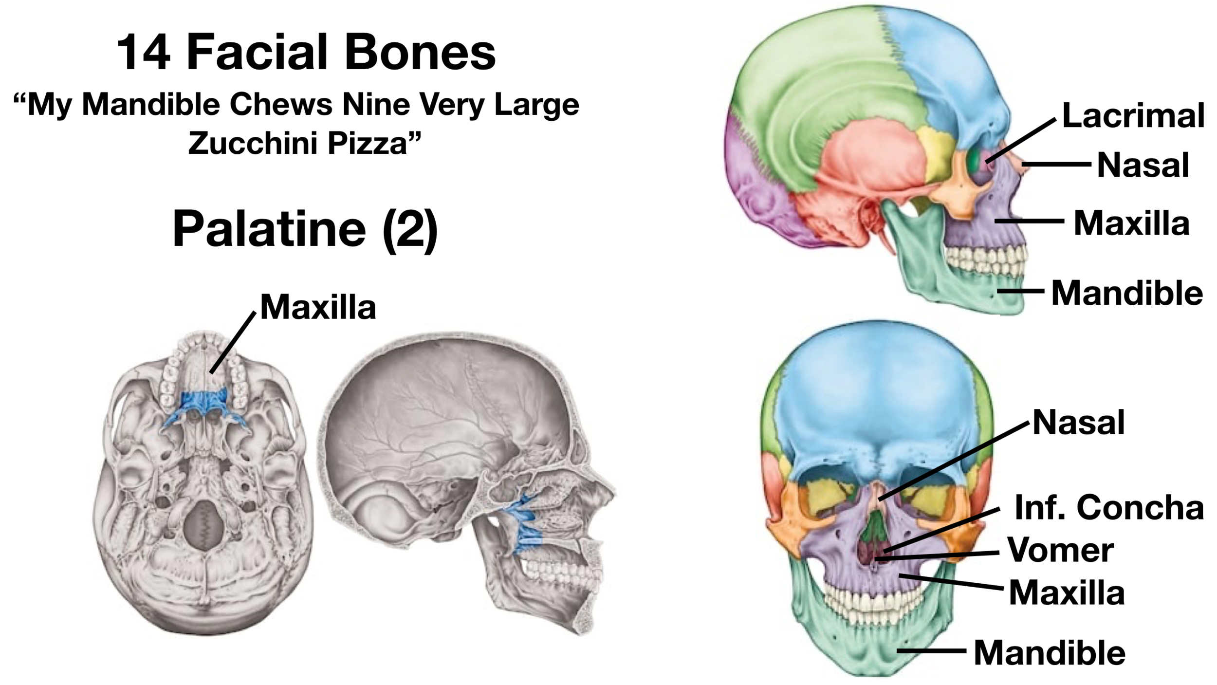
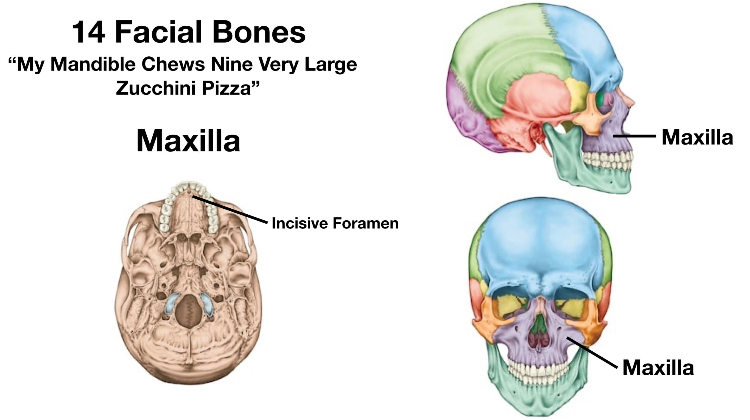
Skull | Definition, Anatomy, & Function | Britannica The facial area includes the zygomatic, or malar, bones (cheekbones), which join with the temporal and maxillary bones to form the zygomatic arch below the eye socket; the palatine bone; and the maxillary, or upper jaw, bones. The nasal cavity is formed by the vomer and the nasal, lachrymal, and turbinate bones.



:watermark(/images/watermark_only_sm.png,0,0,0):watermark(/images/logo_url_sm.png,-10,-10,0):format(jpeg)/images/anatomy_term/skull/eEsfu70EOMx1TlBf5tYAiA_Go0bFvBvzClwSivuaiELg_head_01.png)


:background_color(FFFFFF):format(jpeg)/images/library/11336/labaled_diagram_main_bones_of_skull.jpg)








Komentar
Posting Komentar